How Does Echocardiography Help in the Diagnosis of a Heart Problem?
Además de esto, ORIX le proporciona la posibilidad de obtener información transparente sobre la tramitación de sus peticiones de asistencia por medio de una única plataforma y más información sobre los artículos de OR Technology y su documentación.
 Si bien también tienen la posibilidad de realizarse para estudios de lesiones musculo-esqueléticas y articulares, tendones y ligamentos, lesiones vasculares y viscerales, y en evaluación oncológica.
Las imágenes de rayos X están libres para su visualización en el portátil justo después de la exposición y se tienen la posibilidad de enviar al dueño y expertos veterinarios por correo o servicios en la nube.
En las razas con pecho tipo bull (por servirnos de un ejemplo, Bulldogs, Boston Terriers, Lhasa Apsos), la proporción cardiaca torácica es por norma general mayor que en las etnias de pecho habitual o profundo.
El acolchado terminado da protección contra golpes al descubridor de pantalla plana y al portátil. E inclusive en el caso de un posible fallo de un computador de la granja de PC, es viable proseguir haciendo un trabajo sin problemas y sin tiempo de inacción agregada debido al doble almacenaje estándar. Las imágenes antiguas que llevan mucho más de uno o dos años guardadas se guardan varias veces en ordenadores locales estándar con discos duros comúnes, la llamada granja de ordenadores. Los tiempos de carga de esta una parte del fichero son sólo ligeramente mucho más lentos que los del servidor de imágenes.
Además de esto, con los sistemas de radiografía digital, una cantidad excesiva de exposición fuera del sujeto puede ofrecer sitio a una falsa interpretación de los datos por la parte del algoritmo de reconstrucción y degradar sustancialmente la calidad de la imagen. Si esto sucede, la exposición debe repetirse con una colimación correcta para poder una imagen aceptable. En la mayor parte de las situaciones, el haz de rayos X debe colimarse a ~1 cm fuera de los límites del sujeto para otorgar una calidad de imagen óptima y protección radiológica para el plantel. La colimación correcta del haz de rayos X no puede sustituirse por el uso de la herramienta de recorte de imágenes libre en la mayoría de los sistemas de software usados para generar imágenes digitales. Esta es una herramienta de posprocesamiento y no interfiere a la calidad de la imagen ni a la reconstrucción. Además de esto, esta herramienta jamás debe emplearse para recortar ninguna anatomía del paciente capturada por la exposición inicial y la reconstrucción.
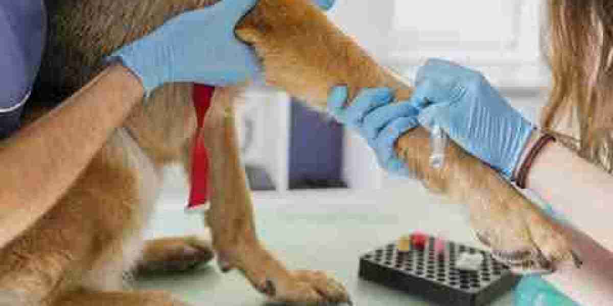
 Abdominal X-rays could additionally be used to help within the diagnosis of conditions involving the intestines, bladder, and different inner stomach organs. A chest X-ray could additionally be beneficial for a pet with a respiratory illness or respiration concern, as nicely as to judge the guts or lungs to help in illness prognosis. We are continually compiling unbiased, practical resources that cowl subjects across the complete breadth of imaging, diagnostics, and therapeutics. This repository of information is built within the form of simply consumable articles and is organized in an on-demand, searchable platform that we name The Knowledge Center. 5,534 pet homeowners requested and obtained a free no-obligation quote from one of many above firms within the final 30 days.
The maximum quantity of knowledge is derived from the radiographic examine when interpretation is done in gentle of the medical and clinicopathologic info available. In this fashion, the more than likely trigger for the animal’s condition can be determined. However, many diseases could cause similar radiographic lesions, and radiographs should be interpreted in light of the entire gestalt of lesions current and clinica veterinaria laboratorio never based mostly on any single lesion if a number of abnormalities are current. Processing algorithms are important to the development of diagnostic images. In many show techniques, the algorithm can be altered to provide enhancement of assorted features of the image.
DICOM Dental Software
In addition, with digital radiography systems, extreme amounts of exposure outside the topic may find yourself in false interpretation of the info by the reconstruction algorithm and substantially degrade image high quality. If this occurs, the publicity must be repeated with correct collimation to attain an acceptable image. In most cases, the x-ray beam ought to be collimated to ~1 cm outdoors the topic limits to offer optimum image high quality and radiation safety for personnel. Proper collimation of the x-ray beam cannot be replaced by use of the imaging cropping device obtainable on most of the software systems used to supply digital pictures.
Head
If the patient is not closely sedated, a workers member sporting the required PPE could additionally be needed to restrain the patient’s head. For sedated patients, a big foam pad can be used to elevate and relaxation the pinnacle and lengthen it away from the forelimb of curiosity. When pulling the top to 1 side, be careful to not rotate the elbow too far medially or laterally. The olecranon should remain centered between the medial and lateral epicondyles of the humerus. Center the beam over the elbow and collimate to incorporate half of the humerus and half of the radius and ulna (FIGURE 41). The marker ought to be placed lateral to the joint indicating which leg is being imaged. A V trough or other positioning system must be used to ensure the affected person is as straight as possible (FIGURE 27).
General Guidelines for Diagnostic Radiography
Our course of includes in-depth industry research into each provider, such as comparing protection options, gathering quotes online to determine pricing and reading evaluations to assess customer support. To better inform our reviews, we’ve surveyed three,000 canine and cat owners nationwide to determine an important parts of pet insurance coverage protection. We’ve additionally purchased pet insurance policy for 10 of our team’s pets to check the client expertise of various suppliers for ourselves. Once all the lesions on the study are recognized, a rational cause for these lesions can be formulated.
Our Diagnostic Imaging Equipment